

Credit score: College of Queensland
Researchers at The College of Queensland have for the primary time captured pictures and video in actual time of early embryonic growth to know extra about congenital delivery defects.
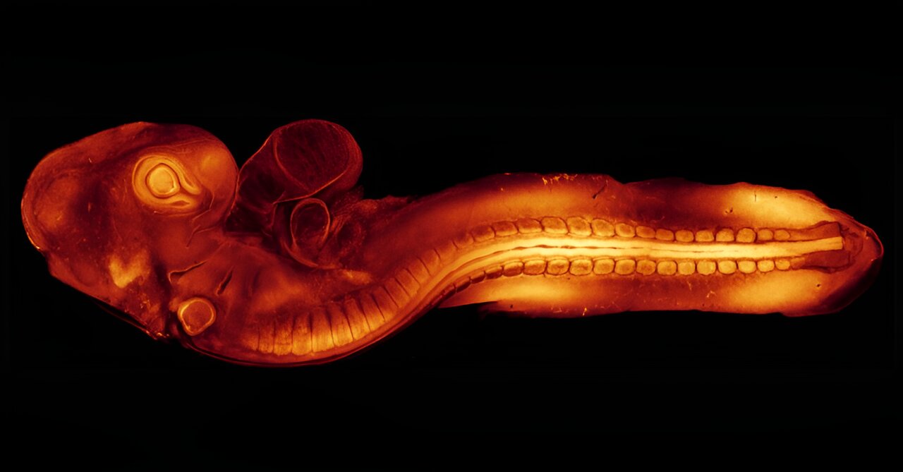
Dr. Melanie White and Dr. Yanina Alvarez from UQ’s Institute for Molecular Bioscience used quail eggs to know how cells start to kind tissues comparable to the guts, mind and spinal twine.
The analysis was revealed within the Journal of Cell Biology by a group that included Marise van der Spuy and Jian Xiong Wang from UQ’s Institute for Molecular Bioscience.
Dr. White mentioned congenital delivery defects have an effect on 3% of Australian infants, with coronary heart defects the most typical and neural tube defects second.
“As a result of quails develop in an egg, they’re very accessible for imaging and their early growth is similar to a human on the time the embryo implants within the uterus,” Dr. White mentioned.
“For the primary time we have now seen high-resolution, real-time imaging of essential early developmental processes.
“Till now, most of our information of post-implantation growth got here from research on static slides, at fastened cut-off dates.”
The IMB researchers have generated quails with a fluorescent protein to disclose the construction, referred to as the actin cytoskeleton, which provides cells form and facilitates motion.
Credit score: College of Queensland
“When cells migrate throughout early growth, they stick out protrusions referred to as lamellipodia and filopodia like arms that attain out and seize onto surfaces permitting the cells to crawl, or attain different cells to convey them nearer collectively,” Dr. White mentioned.
“We had been capable of picture the filopodia from coronary heart stem cells deep contained in the embryo as they first made contact by protruding protrusions and gripping to their environment and one another to kind the early coronary heart.
“It is the primary time anybody has captured the cell’s actin cytoskeleton facilitating this contact in dwell imaging.”
The researchers additionally imaged the open edges of the neural tube and it being ‘zipped up’ to start to kind the mind and spinal twine.
“We noticed how the cells reached throughout the open neural tube with their protrusions to contact the alternative aspect—the extra protrusions the cells shaped, the quicker the tube zipped up,” Dr. White mentioned.
“If this course of goes awry or is disrupted and the tube does not shut correctly in the course of the fourth week of human growth, the embryo can have mind and spinal twine defects.
“Our intention is to search out proteins or genes that may be focused sooner or later or used for screening for congenital delivery defects.
“We’re very excited on the potentialities that this new quail mannequin now provides to review growth in actual time.”
Extra info:
Yanina D. Alvarez et al, A Lifeact-EGFP quail for learning actin dynamics in vivo, Journal of Cell Biology (2024). DOI: 10.1083/jcb.202404066
Offered by
College of Queensland
Quotation:
Quail imaging provides insights into congenital delivery defects (2024, July 1)
retrieved 5 July 2024
from https://phys.org/information/2024-07-quail-imaging-insights-congenital-birth.html
This doc is topic to copyright. Other than any truthful dealing for the aim of personal research or analysis, no
half could also be reproduced with out the written permission. The content material is supplied for info functions solely.


