
To get a more in-depth, real-time take a look at growing fetuses, and to higher perceive the potential causes of delivery defects and different well being points, scientists have turned to a supply you won’t have anticipated: quail eggs.
Actually, our earliest developments as residing beings are much like these of quails, and since their embryos develop inside eggs, they are often scanned comparatively simply. Avian eggs have lengthy been favored by scientists for the examine of embryos.
Right here, researchers in Australia used eggs carrying quails bred to precise a fluorescent peptide that binds to actin proteins that kind the construction of the early embryo, referred to as the actin cytoskeleton. This method allowed them to observe cells migrating and coming collectively to kind organs.
“For the primary time we’ve seen high-resolution, real-time imaging of necessary early developmental processes,” says developmental biologist Melanie White from the College of Queensland.
“Till now, most of our information of post-implantation improvement got here from research on static slides, at mounted cut-off dates.” frameborder=”0″ enable=”accelerometer; autoplay; clipboard-write; encrypted-media; gyroscope; picture-in-picture; web-share” referrerpolicy=”strict-origin-when-cross-origin” allowfullscreen>The group was in a position to see the very early levels of coronary heart, mind, and spinal wire formation. A wide range of microscope devices have been used to seize the fluorescent marker, which outlined the motion of cells.
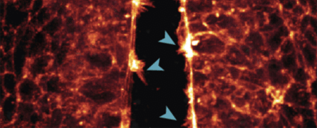
One of many observations made was of the neural tube, the precursor to the central nervous system, being ‘zipped up’ as cells joined collectively.
“We noticed how the cells reached throughout the open neural tube with their protrusions to contact the alternative aspect – the extra protrusions the cells shaped, the sooner the tube zipped up,” White explains.
“If this course of goes awry or is disrupted and the tube would not shut correctly through the fourth week of human improvement, the embryo can have mind and spinal wire defects.”The neural tube ‘zipping up’ throughout early improvement. Blue arrows present cells extending from the perimeters of the neural folds into the open neural tube. (Alvarez et al., The Journal of Cell Biology, 2024)There have been comparable connections made within the stem cells that will finally kind the hearts of the quails.
“We have been in a position to picture the filopodia from coronary heart stem cells deep contained in the embryo as they first made contact by protruding protrusions and gripping to their environment and one another to kind the early coronary heart,” says White.
“It is the primary time anybody has captured the cell’s actin cytoskeleton facilitating this contact in dwell imaging.”
Apart from providing an interesting perception into formative years, the examine is necessary for rising our information of how and why delivery defects happen. When the connection processes fail, that may result in issues for the growing toddler.
Seeing these organic transformations happen in actual time, and on the smallest scales, ought to be helpful sooner or later for mitigating or not less than figuring out the danger of delivery defects. A lot extra research of quail eggs utilizing this course of at the moment are deliberate by the group.
Scientists are persevering with to enhance their fashions and their understanding of what occurs within the womb, and thru that we will work to make extra pregnancies as wholesome as doable.
“Our purpose is to seek out proteins or genes that may be focused sooner or later or used for screening for congenital delivery defects,” says White.
“We’re very excited on the prospects that this new quail mannequin now presents to check improvement in actual time.”The analysis has been revealed in The Journal of Cell Biology.

Unimaginable New Tech Lets Scientists Watch Fetuses Develop in Actual Time : ScienceAlert

- Trending
- Comments
- Latest


